
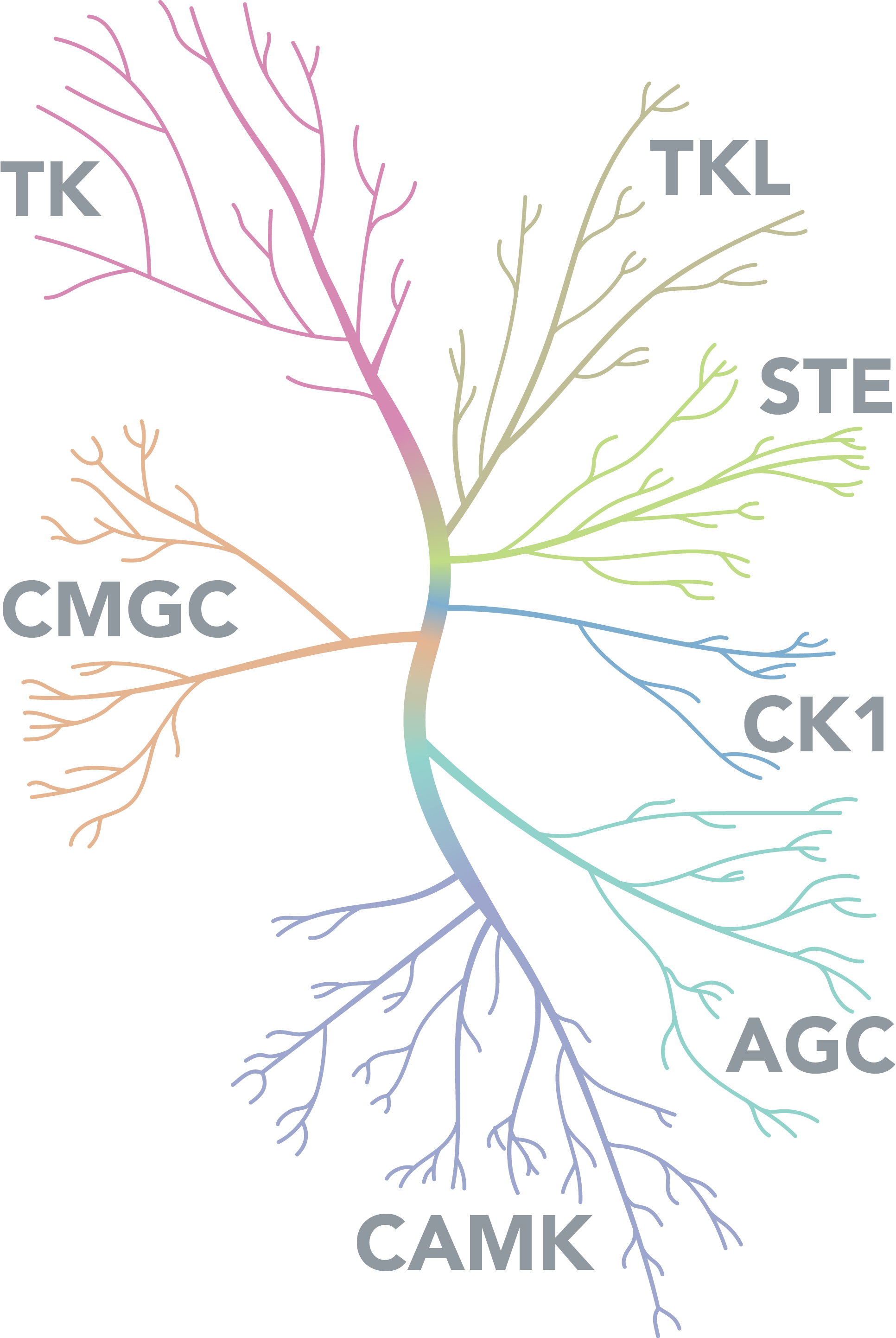
An illustration of the protein kinases that are discussed in this Biology Diagrams The Aurora kinase family share a highly conserved C-terminal catalytic domain and a short N-terminal domain , and function in the regulation of mitosis and cytokinesis . Deregulation of Aurora kinases causes a defect in spindle assembly, checkpoint function, and cell division, leading to chromosome missegregation or polyploidization [ 73 ].

Regulation by Phosphorylation. Activation segment phosphorylation regulates kinase activity of several kinases during mitosis. Both Aurora A and Aurora B kinases undergo auto-phosphorylation within their activation segments (T288 and T248 in Aurora A and Aurora B, respectively) which is essential for their increased kinase activity (Bayliss et al., 2003; Sessa et al., 2005). This review uses specific examples from the field of mitosis to highlight key features of a signalling response that rely on cooperativity between kinases and phosphatases (see Box 1 for an overview). We discuss each of these properties in turn from a systems perspective to rationalise why kinase-phosphatase integration is important, before providing examples where this has been shown to be

Substrates of Mitotic Kinases Biology Diagrams
Dysregulated mitosis can lead to cancer and other diseases. In this issue of Science Signaling, two papers expand our knowledge of the intricate interactions between critical kinases and phosphatases that drive mitotic entry and exit. Polo-like kinase 1 (Plk1) is a serine-threonine kinase that is activated by the kinase Aurora A directly and by Polo-like kinase 1 (PLK1) protects against genome instability by ensuring timely and accurate mitotic cell division, and its activity is tightly regulated throughout the cell cycle. Although the The fact that some mitotic kinase anchoring proteins, such as Cyclins and FAM83D, are quickly turned over following mitotic exit, whereas their interacting kinases are not, not only provides insights into their importance in controlling protein kinase function during mitosis, but suggests that their premature proteolysis may act as an effective
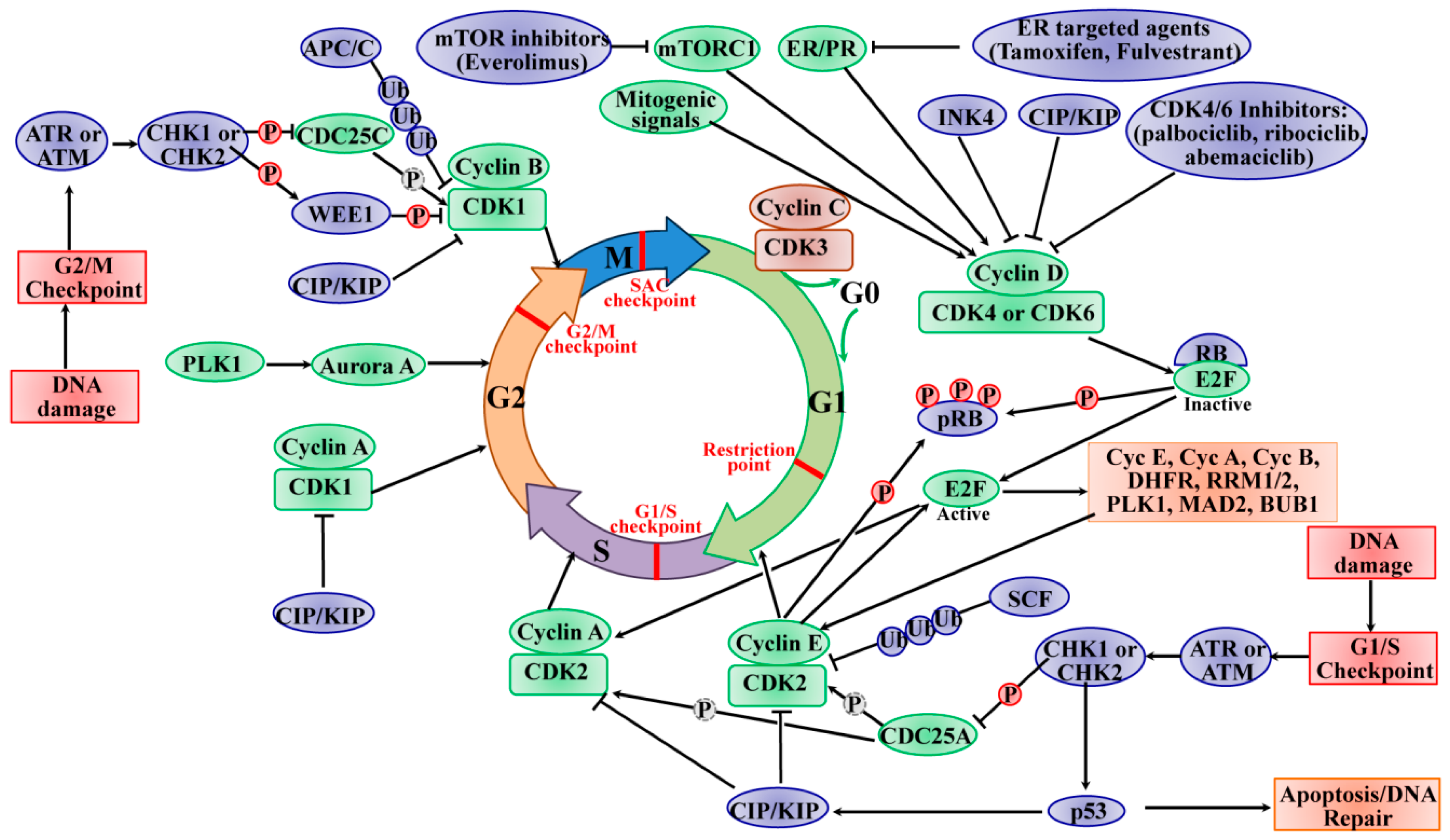
Mitosis is controlled by the kinases: Cdk1/cyclin B, the Aurora family members Aurora A and Aurora B (73% identical in sequence in kinase domain), Polo-like kinase 1 (Plk1), and Nek2. Cdk1/cyclin B is the master regulator with roles in triggering nuclear envelope breakdown, microtubule dynamics, spindle formation, and the separation of
New connections: Kinases and phosphatases in control of mitosis Biology Diagrams
Other than its roles in mitosis, Aurora A kinase was recently found to be involved in autophagy as well. Ahn N.G. Distinct cell cycle timing requirements for extracellular signal-regulated kinase and phosphoinositide 3-kinase signaling pathways in somatic cell mitosis. Mol. Cell. Biol. 2002;22:7226-7241. doi: 10.1128/MCB.22.20.7226-7241.2002. The PLK family comprises five members. PLK1 is a multifunctional kinase implicated in various aspects of mitosis and cytokinesis (11, 28, 29).By contrast, PLK4 functions in S phase as the key centriole assembly-promoting factor, and it is only found in species that have centriolar centrosomes (30-33).PLK2, PLK3, and PLK5 are only present in vertebrates and have diverse functions in
