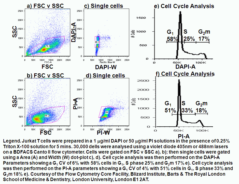
Basic Methods of Cell Cycle Analysis Biology Diagrams If you want to have specific markers, you could detect the cyclins specific for the different phases of the cell cycle by western blot, and correlate that with your BrdU data. Cite 2 Recommendations Cell cycle phases are roughly assigned based on the significance of cell cycle marker gene expressions, with the S phase approximately assigned from 0.5pi to pi, the G2M phase from pi to 1.75pi, the G1 phase from 1.75pi to 0.25pi and the remaining region as undivided . Leveraging the pretrained reference embedding, Tricycle enables rapid cell To examine cell cycle variation in our data, we assign each cell a score, based on its expression of G2/M and S phase markers. An overview of the cell cycle phases is given in the image below: Adapted from Wikipedia (Image License is CC BY-SA 3.0) G0: Quiescence or resting phase. The cell is not actively dividing, which is common for cells that

Phases of the cell cycle. Phases G1, S, and G2 comprise interphases of the cell cycle. M (mitosis) comprises 4 distinct phases: prophase, metaphase, anaphase, and telophase. G0 is the resting phase where cells exist in a quiescent, non-dividing state. Learn immune cell type markers and protocols. The Invitrogen Immunology at Work Resource 3. Cell Cycle and Markers of the Cell Cycle Phases. The cell cycle is composed of four distinct phases. They are the G1 (gap 1), S (DNA synthesis), G2 (gap 2), and M (mitosis) phases . The progression of cells through the cell cycle is controlled by highly orchestrated steps reacting to intracellular and extracellular signals.

Introduction to single Biology Diagrams
Cyclin A/CDK2 are highest at the G2 phase of the cell cycle whereas Cyclin B/CDK1 are highest at the M phase of the cell cycle. I am wondering Is there a good marker(s) for cell cycle arrest Using this marker, we observed changes in the nuclear distribution of Dnmt1 during cell cycle progression. Based on the different nuclear distribution of DNA Ligase I and Dnmt1 in G2 and G1, we demonstrate that the combination of both proteins allows the direct discrimination of all cell cycle phases using either immunostainings or fusions with However, the identification of cell cycle phase marker genes in humans with high resolution is still a challenge. Rapid developments in single-cell technology have made it possible to dissect cells on a large scale with unprecedented resolution. Sequencing for individual cells facilitates our study of gene expression within cells.
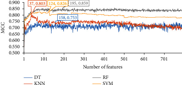
Identification of Human Cell Cycle Phase Markers Based on Single-Cell RNA-Seq Data by Using Machine Learning Methods. This article is part of Special Issue: (G1), DNA synthesis (S), gap 2 (G2), and mitosis (M). Determining which cell cycle phases a cell is in is critical to the research of cancer development and pharmacy for targeting cell