Cell ReproductionMitosis Biology Diagrams The process is longer due to the phases of prophase which takes place in two phases i.e prophase I and prophase II. Prophase I is quite complex which involves the pairing up of the homologous chromosomes and the exchange of genetic information. It defines the difference between mitosis and meiosis. Prophase II is very similar to the mitotic Similar changes were seen in wild-type cells in a previous study. 4 Then, during prophase (−20 to 0 min relative to NEBD) and prometaphase (after NEBD), more cells showed the resolved configuration (4 dots; 2 of each color; pink pattern) and the folded configuration (2 pairs of colocalized red and green dots; red pattern) in both cell lines
+coils+into+chromosomes..jpg)
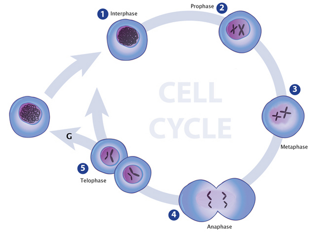
It occurs in several stages, each of which consists of a stereotyped set of changes in cell contents and structure. In this article, we will look at the stages of mitosis and its clinical relevance. To help with this, at the start of prophase, chromatin begins condensing into chromosomes. In addition, mitotic spindles begin to form. phase chromatin changes occur at nuclear pores, on the inner surfaceofthenuclearenvelope, andmoststrikinglyinthenucle-olus. There, proteins involved in rRNA processing move away their proximity to chromatin during prophase. However, about 20% remain relatively unaffected (Figures 3D and 3E; orange cluster). This behavior was well reproduced During prophase, the chromatin condenses into visible chromosomes. Each chromosome consists of two sister chromatids. These are connected at a region called the centromere. The mitotic spindle also begins to form during this stage. Understanding the carbon cycle is essential for addressing climate change and sustaining ecosystems. Through

Nested Irreducible Complexity Biology Diagrams
In prophase, the chromatin condenses into discrete chromosomes. The nuclear envelope breaks down and spindles form at opposite poles of the cell. Prophase (versus interphase) is the first true step of the mitotic process. During prophase, several important changes occur: Chromatin fibers become coiled into chromosomes, with each chromosome
+Centrioles+move+to+poles..jpg)
Prophase. During prophase the nucleoli disappear and the chromatin fibers thicken and shorten to form discrete chromosomes visible with the light microscope. Each replicated chromosome appears as two identical chromatids joined at the centromere.. The chromatids shorten and thicken and become more tightly coiled; the individuality of the separate chromosomes becomes clear. Early prophase chromatin undergoes an orderly transition as proteins leave chromatin in successive waves that form relatively tight clusters in our analysis. We expected that these early events might involve chromatin changes required to shape mitotic chromosomes. Indeed, HMGN1 and HMGA1 are two of the earliest proteins to leave chromatin.