
Clinical Pathways Biology Diagrams Sagittal section of the mouth, nose, pharynx, and larynx. (Reproduced with permission from Anatomy of the Human Body.2) A Symposium: Normal Endoscopic Anatomy/Merati and Rieder August 18, 2003 THE AMERICAN JOURNAL OF MEDICINE Volume 115 (3A) 11S. the oropharynx as well. Normal Endoscopic Anatomy/Merati and Rieder 12S August 18, 2003 THE There are 12 major anatomy systems: Skeletal, Muscular, Cardiovascular, Digestive, Endocrine, Nervous, Respiratory, Immune/Lymphatic, Urinary, Female Reproductive, Male Reproductive, Integumentary. Muscular System The muscular system is responsible for the movement of the human body. Drawing of an endoscope for fetal detection, or "fetoscope" An endoscope is an inspection instrument composed of image sensor, optical lens, light source and mechanical device, which is used to look deep into the body by way of openings such as the mouth or anus.A typical endoscope applies several modern technologies including optics, ergonomics, precision mechanics, electronics, and software
We can make a virtual dissection image of the human body using a virtual dissection simulator and then navigate inside an organ using a virtual endoscope. To improve the navigation performance during virtual endoscopy, our system warns the user about any potential collisions that may occur against the organ's wall by taking the virtual control Endoscopes play an important role in modern medicine, allowing examination of various parts of the human body, including the esophagus, stomach, and small intestine. It acts as a crucial determinant for the diagnosis and treatment of different illnesses. It employs endoscopic scopes, which are advanced devices used to view and examine parts within the body … A Comprehensive Guide of

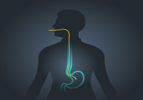
Human Body Diagrams Biology Diagrams
This single-authored book is a review of basic endoscopic anatomy and a presentation of the author's personal observations and recommendations in the technique of endoscopy and interpretation of endoscopic findings. The purpose of the book is to provide basic information to gastrointestinal trainees in their efforts to achieve competence in gastrointestinal endoscopy.

Given that individual fibers can be thinner than human hair, fiber optics is one of the best techniques to enter and view different areas of the body. Endoscope Biopsy -Using this procedure, the physician inserts an endoscope through a body opening or a tiny incision to reach the area of interest. By using biopsy forceps, the physician Fig. 4.1 displays an endoscopic image taken in the sigmoid colon which is the part of the large intestine that is closest to the rectum. Figure 4.1. The orientation and thereby the navigation within the human body depends on the experience of the surgeon. Due to the lack of intuitive visual comparison to the environment, the narrow field of It employs endoscopic scopes, which are advanced devices used to view and examine parts within the body through its normal apertures. In this article, we take a closer look at endoscopes, analyzing their anatomy, functions, and reasons why routine maintenance is necessary and replacement should be done periodically. The Anatomy of Endoscopy System
Virtual Anatomical and Endoscopic Exploration Method of Internal Human ... Biology Diagrams
Medical Art Library is a resource for teachers, students, health professionals or anyone interested in learning about the anatomy of the human body. We are medical artists who love anatomy. We believe that Illustrations can help you focus on key structures, see relationships, and quickly understand anatomy- in a way that words alone can't.