Exploring the Differences of Cytokinesis in Plant and Animal Cells Biology Diagrams Learn about cytokinesis, the final step of cell division in eukaryotes, and how it differs in animal and plant cells. Find out the stages, mechanisms, and examples of cytokinesis with a diagram and FAQs.

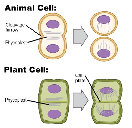
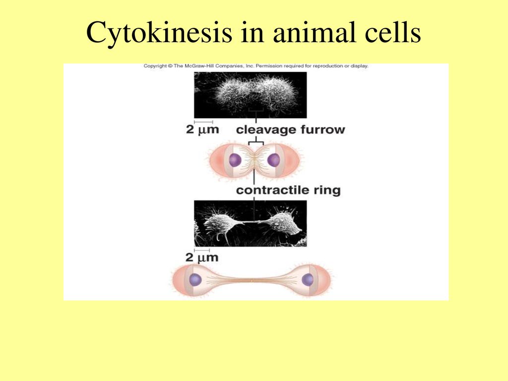
In animal cells, cytokinesis is achieved when a contractile ring of the cell microtubules form a cleavage furrow that divides the cell membrane into half. The microtubules used during cytokinesis are those generated during the initial stages of division and they contribute to the restructuring of the new cell.

Cytokinesis Biology Diagrams
The cell cycle culminates in the division of the cytoplasm by cytokinesis. In a typical cell, cytokinesis accompanies every mitosis, although some cells, such as Drosophila embryos (discussed later) and vertebrate osteoclasts (discussed in Chapter 22), undergo mitosis without cytokinesis and become multinucleate. Cytokinesis begins in anaphase and ends in telophase, reaching completion as the Explore the mechanisms of cytokinesis in animal cells, focusing on contractile ring dynamics, membrane ingression, and the regulation of cell division completion. BiologyInsights Team. Published Mar 11, 2025. Cell division ensures the proper distribution of genetic material and cellular components into daughter cells. In animal cells

Cytokinesis is the final step of cell division during which the two daughter cells become physically separated. It begins right after chromosome segregation in anaphase, when a cytokinetic or cleavage furrow forms at the equatorial cortex and ingresses inward to bisect the mother cell, and terminates with the physical detachment of the two daughter cells (). Cytokinesis is the final step of eukaryotic cell division that separates the cytoplasm and organelles of two daughter cells. Learn how cytokinesis occurs in animal and plant cells, and see diagrams and quizzes to test your knowledge.
Cytokinesis: In Animal and Plant Cells Biology Diagrams
Cytokinesis is the part of cell division when the cytoplasm of a single eukaryotic cell divides into two daughter cells. Learn about the steps, mechanisms and differences of cytokinesis in animal and plant cells, and the role of the central spindle and actin-myosin ring. Cytokinesis in animal cells can be categorized into three major steps: 1) cleavage plane specification, 2) formation and maintenance of the actomyosin ring, 3) constriction of the ring to produce two daughter cells. Although multiple mechanisms of cleavage plane positioning have been proposed, central to all these mechanisms are MTs. Cytokinesis, the final step in cell division, partitions the contents of a single cell into two. In animal cells, cytokinesis occurs through cortical remodeling orchestrated by the anaphase spindle. Cytokinesis relies on a tight interplay between signaling and cellular mechanics and has attracted the attention of both biologists and physicists

Cytokinesis is one of the most dramatic changes in cell shape and requires an extensive reorganization of the cell's cytoskeleton. Here, we describe the cytoskeletal structures, factors, and signaling pathways that orchestrate this robust and yet highly dynamic process in animal cells.