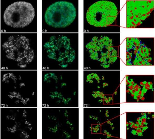
Fluorescence lifetime imaging to study DNA compaction and gene activities Biology Diagrams Fluorescence microscopy is one of the most important approaches in the cell biologist's toolbox for studying the mitotic spindle. In fact, many of the key insights into our understanding of mitosis have been enabled by the visualization of mitotic processes using fluorescence microscopy. Here, we su … Mitosis is a highly dynamic and choreographed process in which chromosomes are captured by the mitotic spindle and physically segregated into the two daughter cells to ensure faithful transmission of the genetic material. Live-cell fluorescence microscopy enables these dynamics to be analyzed over diverse temporal scales.
73.0K Views. Mitosis is a form of cell division in which a cell's genetic material is divided equally between two daughter cells. Mitosis can be broken down into six phases, during each of which the cell's components, such as its chromosomes, show visually distinct characteristics. Advances in fluorescence live cell imaging have allowed scientists to study this process in great detail
Live fluorescence imaging of chromosome segregation in cultured cells Biology Diagrams
Both LSCM and SDCM systems are available with multiple lasers for imaging a range of fluorescent probes. A third alternative for imaging mitosis is to use a wide-field fluorescence microscope and perform deconvolution to remove out-of-focus fluorescence . Turnkey systems are available for this mode of microscopy (e.g. DeltaVision). 1.1 Overview of Fluorescence Imaging And Mitosis. In the late 1800's Flemming, Van Beneden and their contemporaries carried out the first studies of chromosome behavior in dividing cells in both living and fixed tissue using the available microscope methodology (reviewed in ). Since that time, tremendous advances in microscope technology and Digital imaging with fluorescence microscopy is becoming a powerful tool to assist scientists in understanding the complex process of mitosis on both a structural and functional level. Fluorescent probes can be attached to specific subcellular components, such as the chromosomes and microtubules, for visualization of mitosis using standard epi

Mitosis is a highly dynamic and choreographed process in which chromosomes are captured by the mitotic spindle and physically segregated into the two daughter cells to ensure faithful transmission of the genetic material. Live-cell fluorescence microscopy enables these dynamics to be analyzed over diverse temporal scales. The impact of fluorescence microscopy on cell biology in general, and on the field of mitosis in particular, is profound. This major impact has occurred due to the confluence of the development of fluorescent reporter molecules that can be delivered to and expressed in live cells and advances in equipment used for imaging. 1.1. OVERVIEW OF FLUORESCENCE IMAGING AND MITOSIS. In the late 1800's Flemming, Van Beneden and their contemporaries carried out the first studies of chromosome behavior in dividing cells in both living and fixed tissue using the available microscope methodology . Since that time tremendous advances in microscope technology and fluorescence

Using Fluorescence Microscopy to Study Mitosis Biology Diagrams
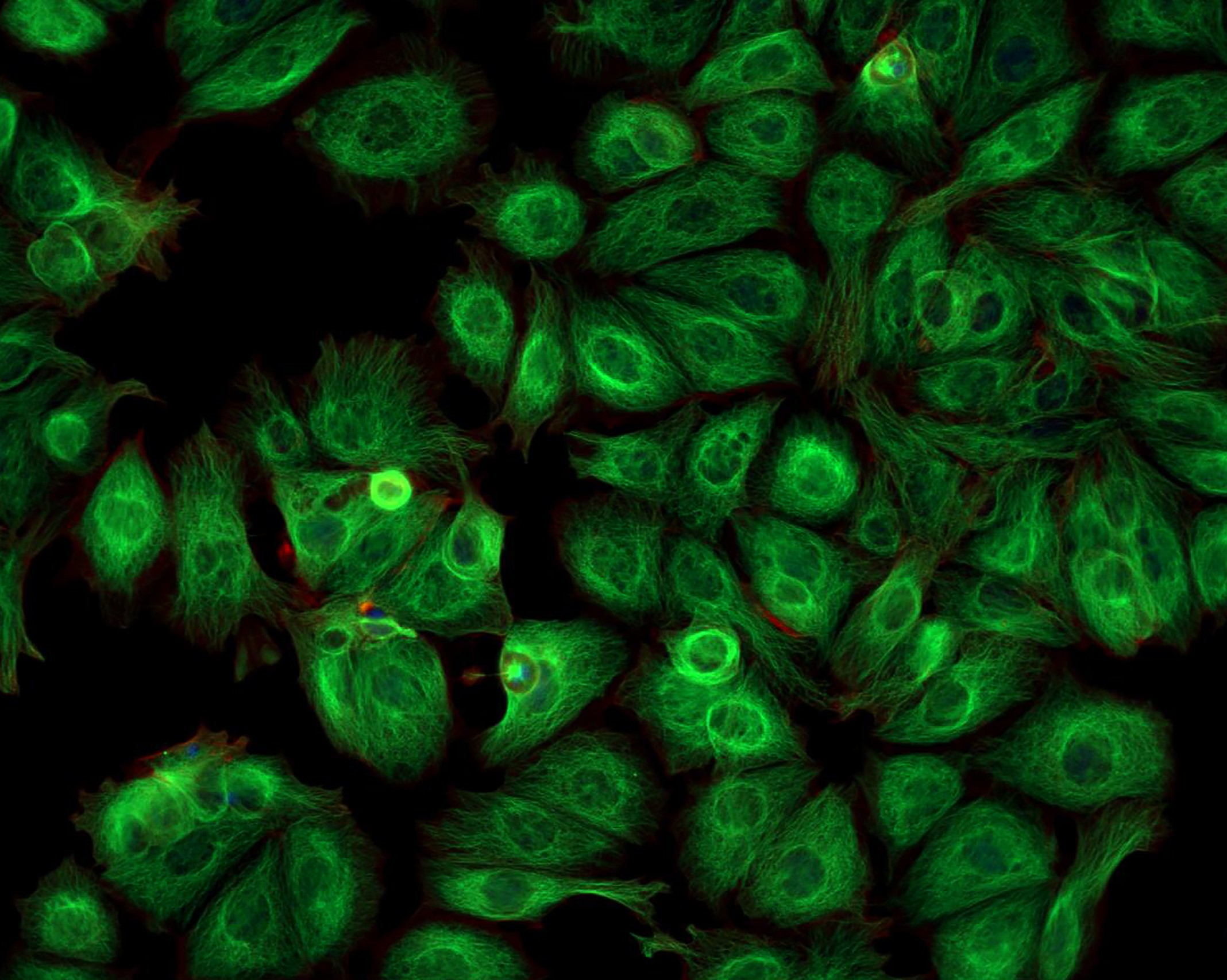
By creating fluorescent fusion proteins varying in wavelength emission, one can further determine if proteins of interest co-localize or concentrate in specific subcellular compartments during mitosis. For use in wide field fluorescence imaging assays described herein, a human cell line was employed that expresses histone H2B fused to green