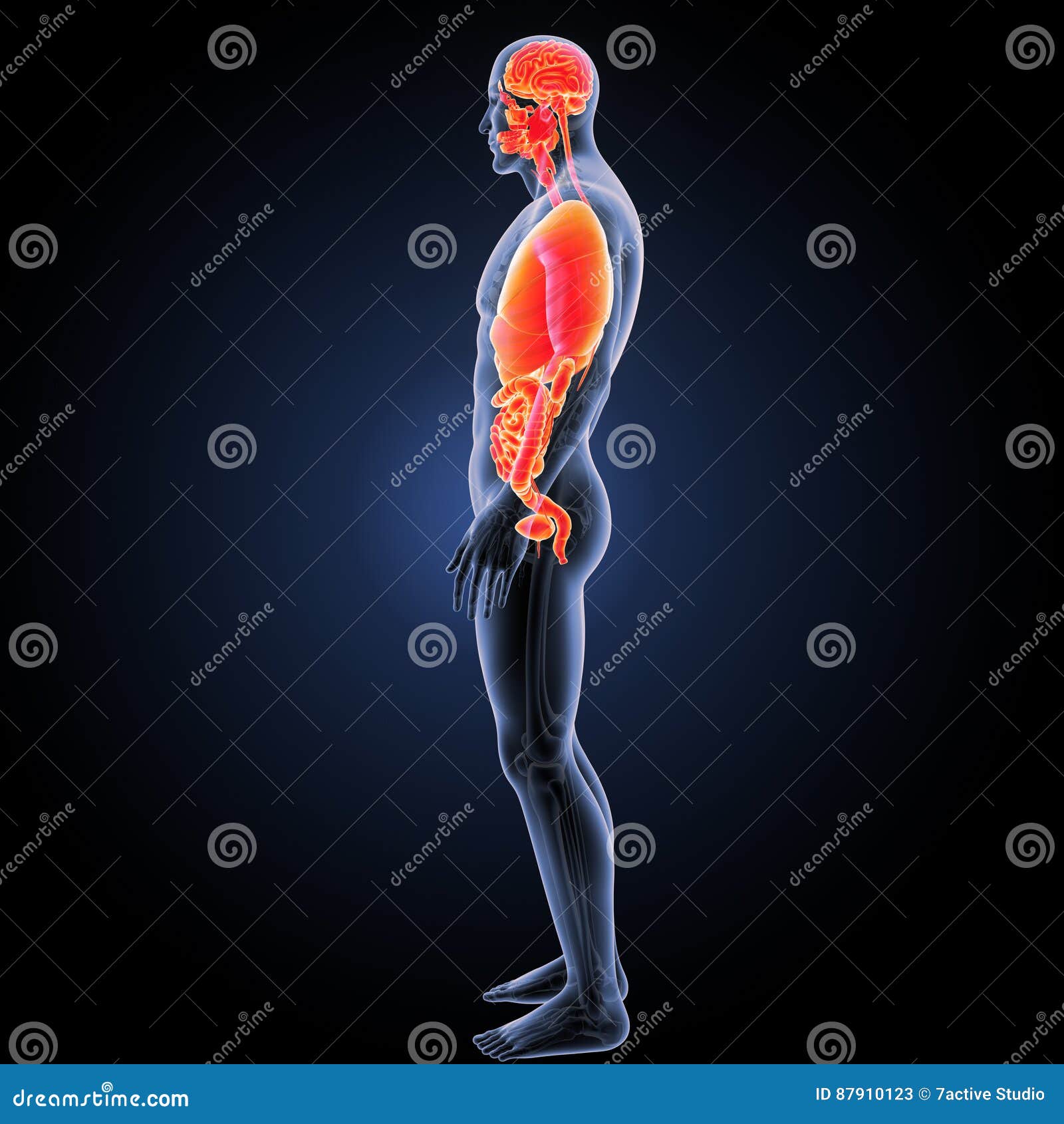
Human Organs with Skeleton Lateral View Stock Illustration Biology Diagrams The lateral view of the human skull provides essential insights into the complex arrangement of bones, sutures, and anatomical landmarks critical for medical diagnosis and surgical planning. This perspective reveals key structures involved in cranial development, sensory function, and mastication.

The bone forms the lateral part of the orbital socket and the floor of the orbit as well. Incisive foramen The incisive foramen is a funnel-shaped opening also known as the anterior palatine foramen. It is found in the hard palate directly behind the incisor teeth. That foramen can be sometimes seen from the posterior view of the skull.

Lateral aspect of cranium Biology Diagrams
The human skull is a fascinating anatomical structure composed of multiple bones that protect the brain and sensory organs while facilitating essential functions like eating and breathing. This detailed anatomical diagram presents both frontal and lateral views of the skull, highlighting 29 distinct anatomical features. Medial and Lateral. Imagine a line in the sagittal plane, splitting the right and left halves evenly. This is the midline. Medial means towards the midline, lateral means away from the midline. Examples: The eye is lateral to the nose. The nose is medial to the ears. The brachial artery lies medial to the biceps tendon. The side view of the skull, known as the lateral aspect of cranium or norma lateralis, shows how the skull appears from the side. In this view, you can see various bones, including those from the neurocranium such as the frontal, parietal, sphenoid (greater wing), temporal, and occipital bones. Bones from the facial skeleton are also visible in this side view, including the nasal, maxilla

The human skull skeletal anatomy lateral view with label. Anatomy Note-November 23, 2024. The human skull is a remarkable anatomical structure, with its lateral view revealing crucial bones and features essential for protecting the brain and facilitating vital functions. This detailed illustration highlights nine key components of the skull's Frank H.Netter MD: Atlas of Human Anatomy, 5th Edition, Elsevier Saunders, Chapter 1 Head and Neck, Skull: Lateral View, Page 6 to 9 and 15. Lateral View, Page 6 to 9 and 15. Chummy S.Sinnatamby: Last's Anatomy Regional and Applied, 12th Edition, Churchill Livingstone Elsevier, Chapter 8 Osteology of the Skull and Hyoid bone, Page 504 and

Open Educational Resources Biology Diagrams
This online quiz is called Human cranial and facial bones lateral view. It was created by member Julie Duncan and has 18 questions. Anatomy of the Human Heart - Internal Structures. Medicine. English. Creator. orkide1 +1. Quiz Type. Image Quiz. Value. 24 points. Likes. 631. Played. 244,409 times. Human Skull Bones (Lateral View) Illustrations Menu. Background Info. The skull (cranium) is the skeletal structure of the head that supports the face and protects the brain. It is subdivided into the facial bones and the cranial bones. The facial bones underlie the facial structures, form the nasal cavity, enclose the eyeballs, and support the Figure 7.3.3 - Lateral View and Sagittal Section of Skull: (a) Lateral View of Skull. The lateral skull shows the large rounded brain case, zygomatic arch, and the upper and lower jaws. The zygomatic arch is formed jointly by the zygomatic process of the temporal bone and the temporal process of the zygomatic bone.

