
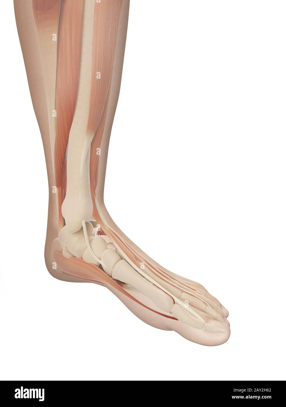
muscular foot anatomy Stock Photo Biology Diagrams These muscles contract to plantar flex the foot --- such as when standing on your tiptoes --- and flex the toes. Shin muscles, such as the tibialis anterior and extensor digitorum longus, dorsiflex the foot and extend the toes. The muscles of the calf also work subtly to stabilize the ankle joint and foot and to maintain the body's balance. Learn about the extrinsic and intrinsic muscles of the foot, their origins, insertions, actions, and nerve supply. The web page covers the dorsal and plantar aspects of the foot, with diagrams and descriptions of each muscle.
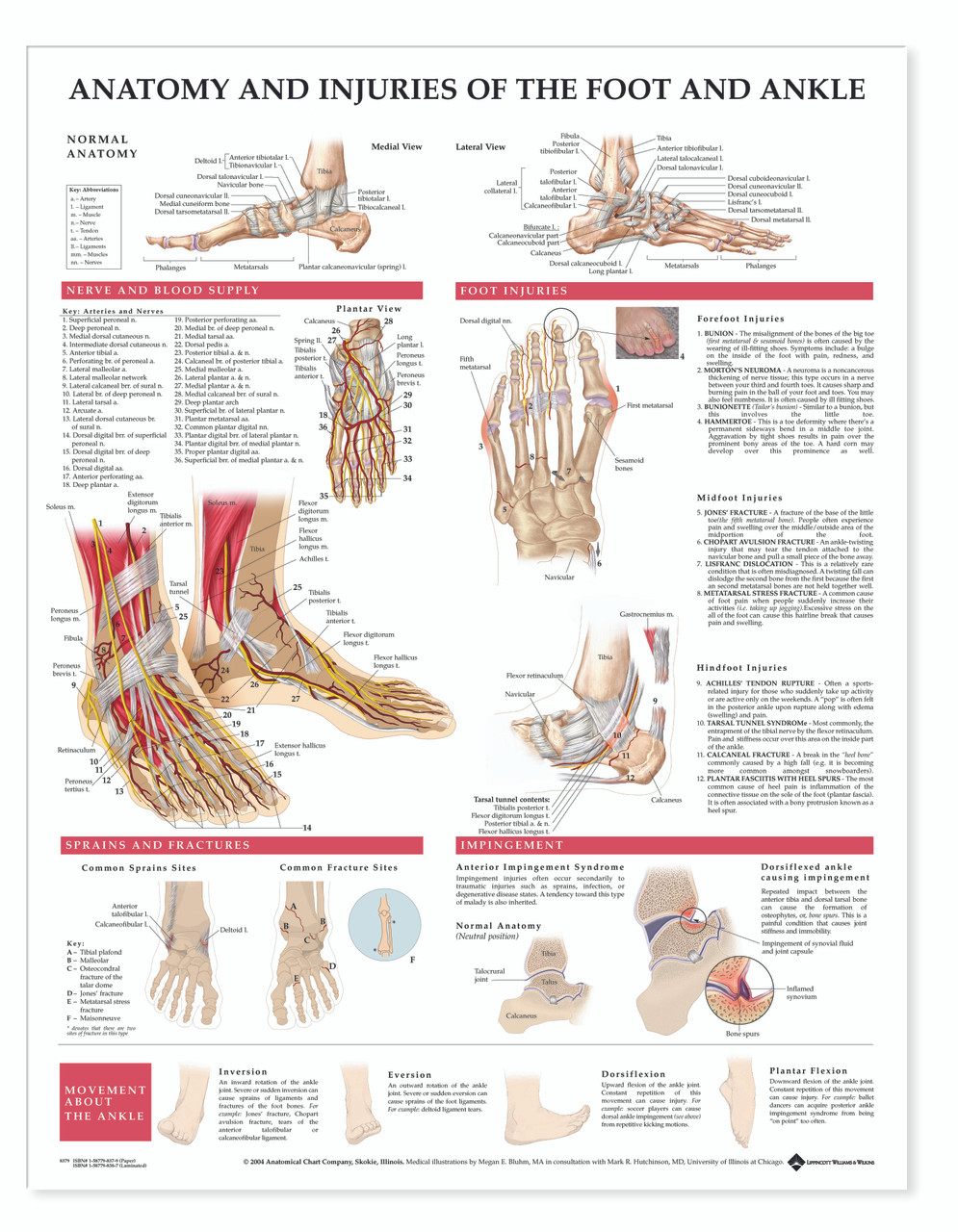
Learn about the complex structure and function of the human foot, which contains 26 bones, 33 joints, and over 100 moving parts. Find out how the foot supports your weight, allows for locomotion, and can be affected by various injuries and disorders.
Easy Anatomy 3D Biology Diagrams
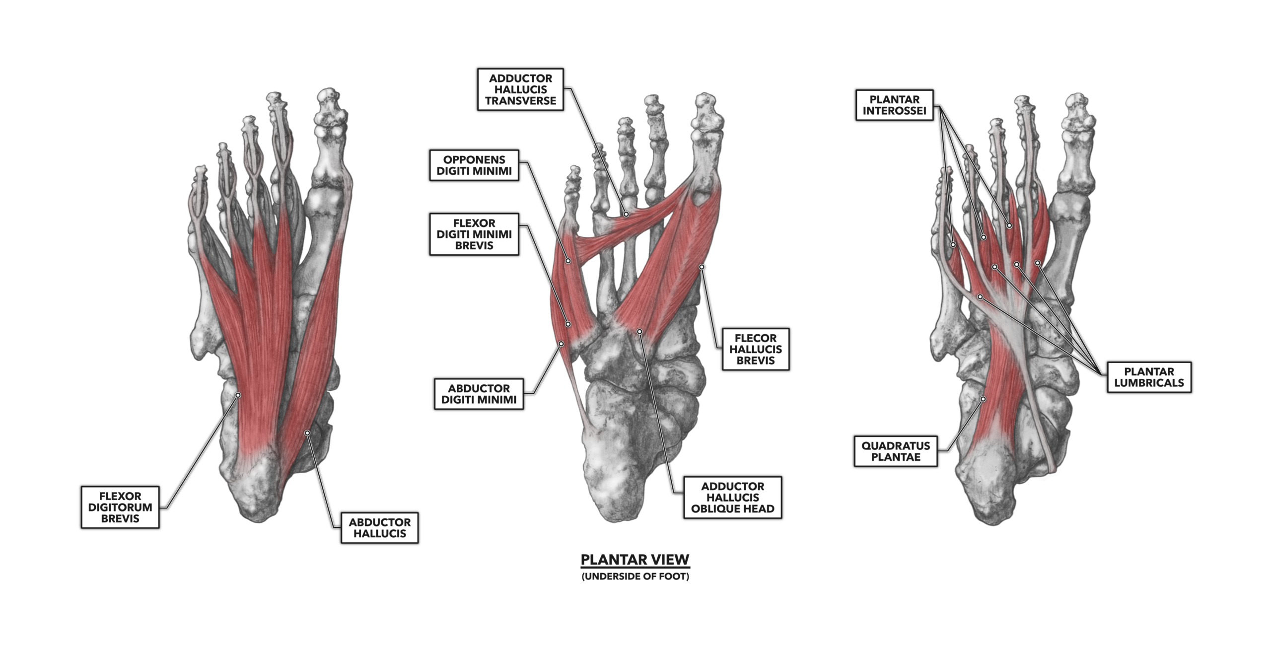
Learn about the anatomy of the ankle and foot, including the bones, joints, ligaments, and muscles. Find out how the foot differs from the hand and how it supports body weight and movement. The extrinsic muscles of the foot originate in the lower leg, whilst the intrinsic muscles are contained within the foot itself. The intrinsic foot muscles act to stabilise the foot and support the arches, as well as to produce fine movement of the toes. The intrinsic foot muscles can be divided into two main groups, plantar and dorsal. The 10 extrinsic muscles of the foot cross the ankle joint to connect with the leg. Tendons and ligaments of the foot Ligaments attach bones to other bones at joints, and there are more than 30 in

There are 29 muscles associated with the human foot: 10 originate outside the foot but cross the ankle joint to act on the foot, and 19 are intrinsic foot muscles. The foot is crucial to human locomotion and postural stability, and the muscles associated with the foot are therefore involved principally in this function. The muscles are aided by the plantar fascia, shaping the posture, shape

Anatomy, Bony Pelvis and Lower Limb, Foot Muscles Biology Diagrams
Learn the anatomy, function, blood supply and innervation of the muscles of the foot. See 3D images and diagrams of the dorsal and plantar muscles of the foot, with detailed descriptions and labels.
The muscles of the dorsum of the foot are a group of two muscles, which together represent the dorsal foot musculature. They are named extensor digitorum brevis and extensor hallucis brevis. The muscles lie within a flat fascia on the dorsum of the foot (fascia dorsalis pedis) and are innervated by the deep fibular or peroneal nerve.
