
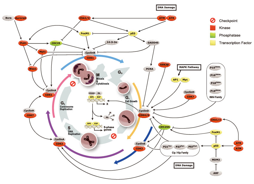
New Insights into CDK Regulators Novel Opportunities for Cancer Biology Diagrams The tumour suppressor protein p53 is a sequence‐specific DNA‐binding protein, that is able to induce either cell cycle arrest or apoptosis at the cell cycle checkpoints. The p53 tumour suppressor gene was first discovered in SV40 transformed cells by the finding that its protein product p53 was tightly bound to the SV40 large T oncogene

Cancer is characterized by uncontrolled tumour cell proliferation resulting from aberrant activity of various cell cycle proteins. Therefore, cell cycle regulators are considered attractive targets in cancer therapy. Intriguingly, animal models demonstrate that some of these proteins are not essenti … Many cell cycle proteins are overexpressed or overactive in human cancers, in particular, D-type and E-type cyclins, cyclin-dependent kinases (CDK4, CDK6 and CDK2), Polo-like kinase 1 (PLK1) and

10.3C: Regulator Molecules of the Cell Cycle Biology Diagrams
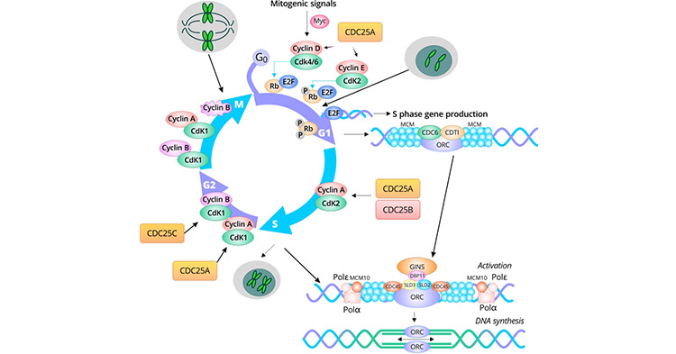
The different cyclins and Cdks bind at specific points in the cell cycle and thus regulate different checkpoints. Figure \(\PageIndex{1}\): Activation of Cdks: Cyclin-dependent kinases (Cdks) are protein kinases that, when fully activated, can phosphorylate and activate other proteins that advance the cell cycle past a checkpoint.
The E2F-dependent transcriptional network includes many genes that encode key proteins in cell cycle and DNA replication control but also genome protection mechanisms and growth. E2F-dependent
Cell-cycle proteins - Latest research and news Biology Diagrams
Key Regulatory Proteins. Cell cycle progression is controlled by regulatory proteins that act as molecular switches, responding to internal and external signals to either promote or halt division. Among the most significant regulators are cyclins, cyclin-dependent kinases (CDKs), and cell cycle inhibitors. Cyclins A cell cycle regulatory protein, p16, is a surrogate marker for HPV infection that reflects the activation of E6- or E7-driven carcinogenesis through degradation of p53 and dysregulation of Rb. A positive result on p16 immunohistochemistry is defined as strong and diffuse nuclear and cytoplasmic staining (known as block staining).
