Optic nerve anatomy Royalty Free Vector Image Biology Diagrams Download scientific diagram | Schematic of the optic nerve and its different segments. Schematic of the optic nerve and its different segments. from publication: A Narrative Review of Point of The optic nerve is a crucial component of the visual system, serving as the primary conduit for transmitting visual information from the retina to the brain. This nerve, also known as cranial nerve II, is composed of approximately 1.2 million nerve fibers, which are the axons of retinal ganglion cells. These fibers bundle together to form the Optic nerve anatomy. The optic nerve is located at the very back of the eye, attached to the retina. Because of its function, the optic nerve is considered part of the nervous system, even though it's located in the eye. The Optic Nerve (cranial nerve II) Nerve (ganglionic) cells as well as millions of nerve fibers make up the optic nerve.
Optic nerve in a cadaver. It is the most prominent neural structure of the orbit. Just before it enters the optic canal, CN II can be found adjacent to CN III, CN VI and the nasociliary nerve, and superomedial to the ophthalmic artery.Within the canal, there are numerous fibrous dural attachments that extend to the pia mater. Learn about the optic nerve, a paired cranial nerve that transmits visual information from the retina to the brain. See the anatomy, function and clinical significance of the optic nerve, as well as its relation to the visual cortex and the eye's blind spot. The optic nerve has been studied heavily because it is a direct extension of the brain. Anatomy . The optic nerve is mainly made up of the axons (nerve fibers) of the retinal ganglion cells from the retina. The optic disc or nerve head is the point where the axons from the retinal ganglion cells leave the eye.
All About Vision Biology Diagrams
The optic nerve also plays a role in controlling the size of the pupil, the small opening in the center of the iris that regulates the amount of light entering the eye. When the pupil constricts, or becomes smaller, it helps to reduce the amount of light entering the eye, which can be beneficial in bright light conditions. [2] Conversely, when the pupil dilates, or becomes larger, it allows In the center of the retina is the optic nerve, a circular to oval white area measuring about 2 x 1.5 mm across. A simplistic wiring diagram of the retina emphasizes only the sensory photoreceptors and the ganglion cells with a few interneurons connecting the two cell types such as seen in Figure 2. Fig. 2. Simple organization of the retina. Learn about the optic nerve, the connection between your eyes and your brain that lets you see. Find out how it works, where it is, and what can affect it.
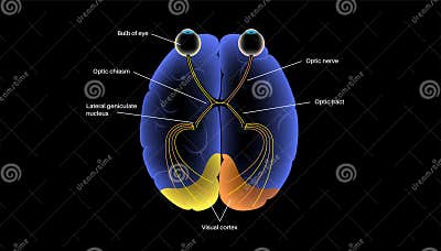
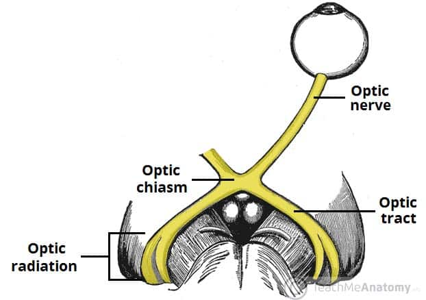
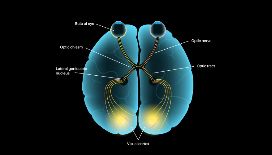
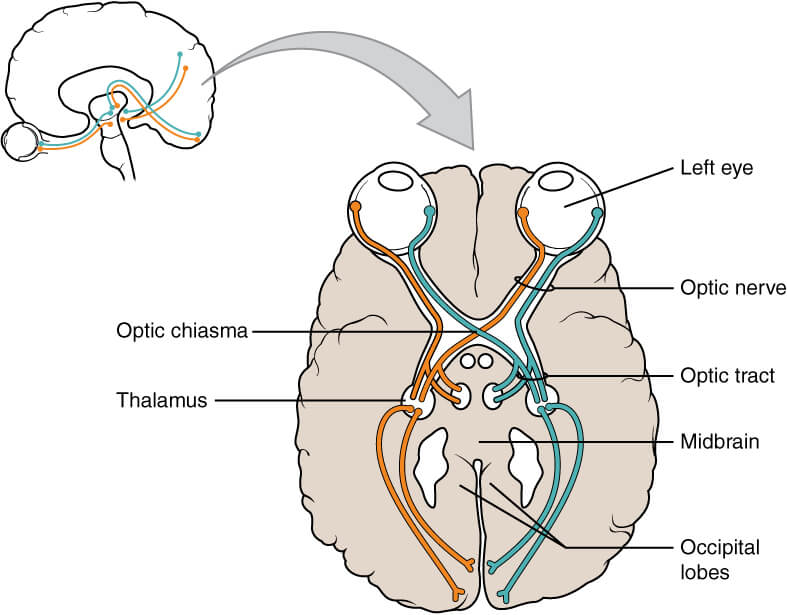
Learn about the optic nerve (CN II), the second cranial nerve that transmits visual information from the eye to the brain. See the anatomical course, the optic chiasm, the optic tract and the optic radiation, and their clinical relevance.