
Skeletal System Diagrams to Label and Color with Reference Information Biology Diagrams Medical Art Library is a resource for teachers, students, health professionals or anyone interested in learning about the anatomy of the human body. We are medical artists who love anatomy. We believe that Illustrations can help you focus on key structures, see relationships, and quickly understand anatomy- in a way that words alone can't.

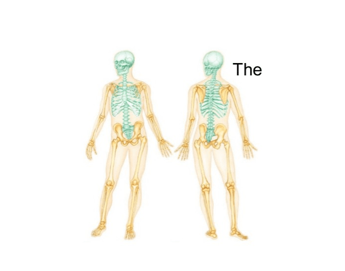
The Skeletal System has many important functions: • Providing support for the body • Storing minerals (calcium, phosphate) • Producing red blood cells • Protecting the organs and tissues • Allowing movement (the bones act as levers) The skeleton can be subcategorized into two divisions: The Axial Skeleton (left, in blue) The human skeletal system is made up of more than 200 bones and has two main parts: the axial and appendicular skeleton. Find labeled diagrams here.
Printable Human Skeleton Diagram Biology Diagrams
The ilium is the big bone of the hip, the ischium is the bone on which one sits and the pubis forms the lower frontal hip bone as seen in the diagram. Femur. The longest and the strongest bone in the human skeletal system as you can observe in the labeled skeleton diagram of the human body. The femur or the thigh bone is closest to the body. An in-depth review of the human skeletal system and its different parts and bones, featuring the beautiful GetBodySmart diagrams and illustrations. Learn the skeletal system anatomy with our tutorials and interactive diagrams below, and discover the bones of the body using labeled worksheets. Learn anatomy faster and remember everything you
Finally, the skeleton grows throughout childhood and provides a framework for the rest of the body to grow along with it. Skeletal System Anatomy. The skeletal system in an adult body is made up of 206 individual bones. These bones are arranged into two major divisions: the axial skeleton and the appendicular skeleton. The axial skeleton runs Given below is a labeled diagram, and tips to help you draw and memorize the names of different parts. Human Skeleton Diagram. Here is a detailed diagram which shows the various bones present in an adult skeletal system. There is a little difference between the male and female skeleton, but for diagrams mostly a male skeletal system is considered. If you need to review the human skeleton for an upcoming test or quiz, this page provides several free human skeleton diagrams to help you study. There are 6 worksheets to choose from. The first two show the labeled human skeleton. One is in color, and the other is in black and white. Finally, the last set of printables show a blank human skeleton.

Human Body Diagrams Biology Diagrams
There are 12 major anatomy systems: Skeletal, Muscular, Cardiovascular, Digestive, Endocrine, Nervous, Respiratory, Immune/Lymphatic, Urinary, Female Reproductive human skeleton, the internal skeleton that serves as a framework for the body. This framework consists of many individual bones and cartilages.There also are bands of fibrous connective tissue—the ligaments and the tendons—in intimate relationship with the parts of the skeleton. This article is concerned primarily with the gross structure and the function of the skeleton of the normal
