The Pelvic Floor Needs New PR Biology Diagrams Learn about the muscles that form the lower limit of the true pelvis and support the pelvic organs. Find out their attachments, blood supply, innervation, function, embryology, and clinical significance.

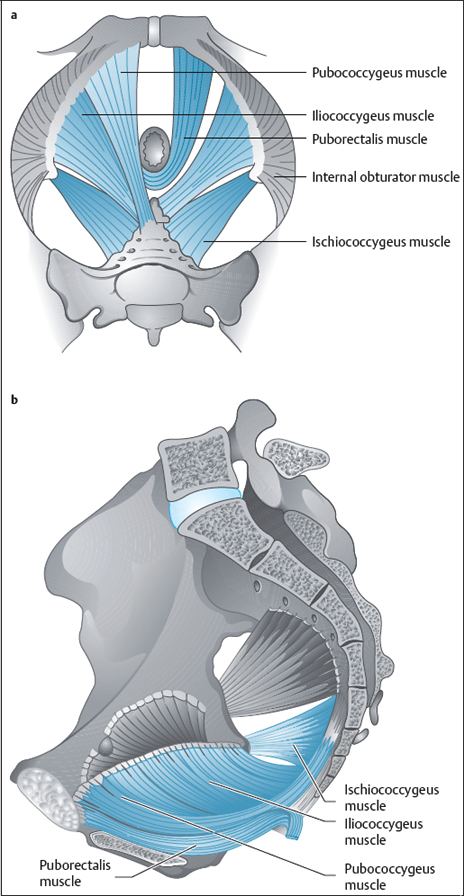
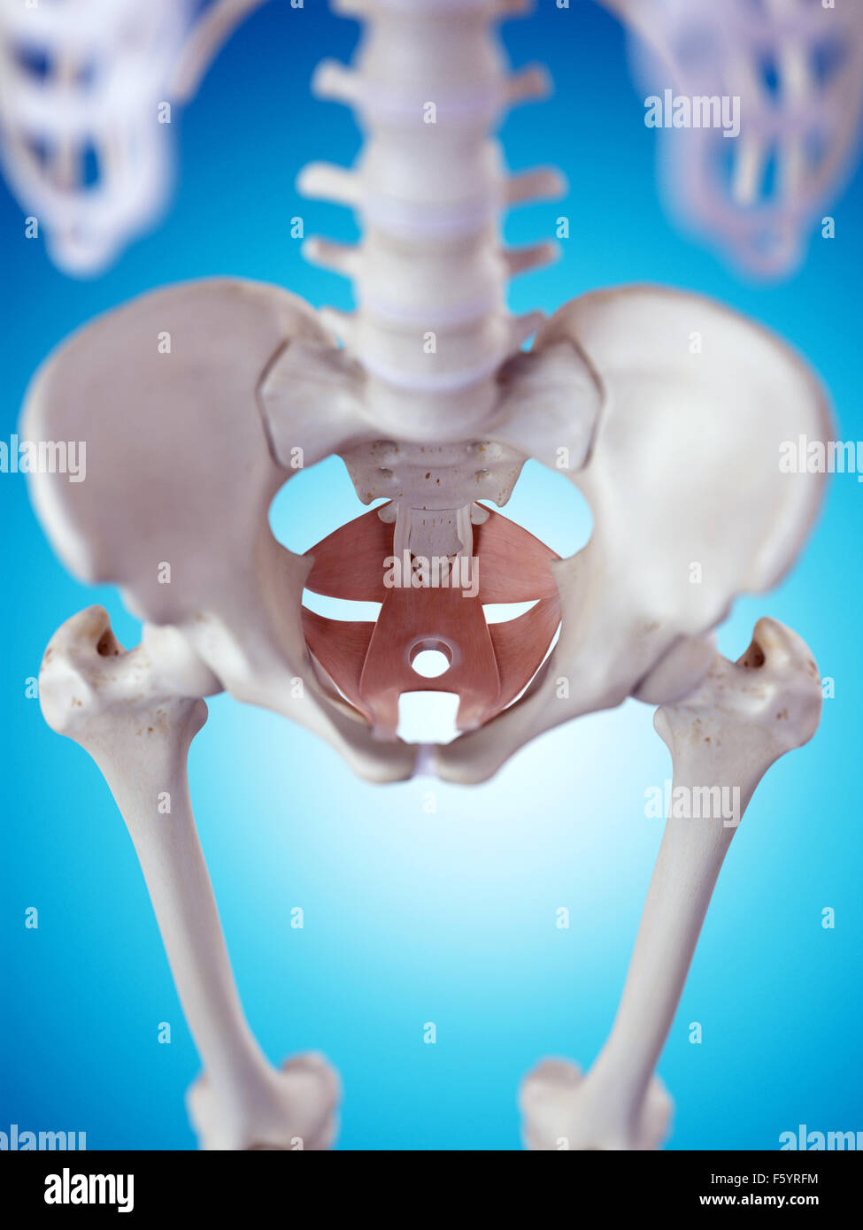
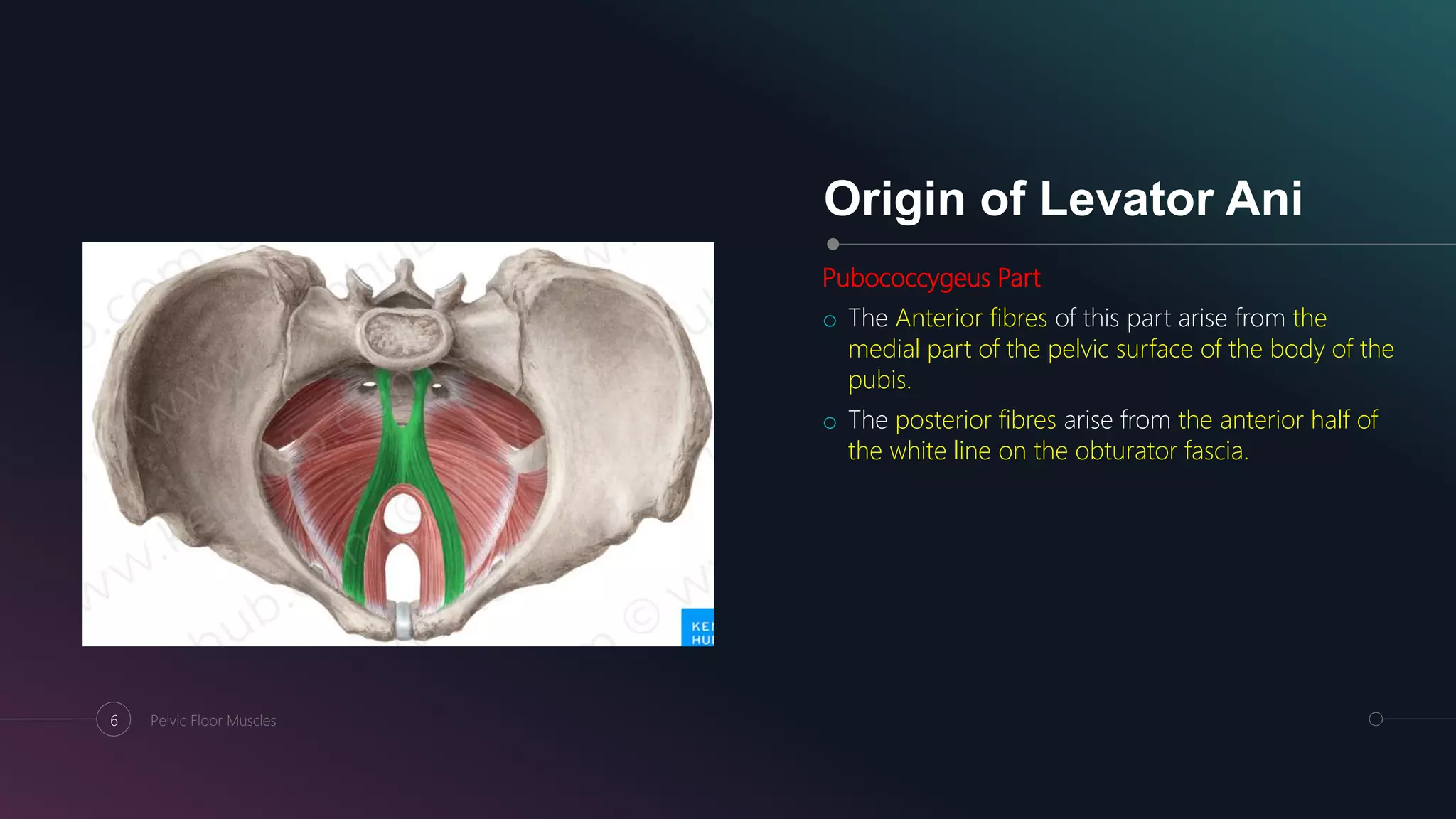
Deep Layer - Pelvic Diaphragm [edit | edit source]. The deepest layer of the pelvic floor muscles is known as the pelvic diaphragm (see Figure 4). It is a broad, funnel-shaped sling of fascia and muscle suspended from bony anchor points in the lesser pelvis (i.e. the area of the pelvic cavity below the linea terminalis).. The muscles of the pelvic diaphragm are:
Overview of Female Pelvic Floor Muscle Anatomy and Physiology Biology Diagrams
Pelvic floor muscle training versus no treatment, or inactive control treatments, for urinary incontinence in women: a cochrane systematic review abridged republication. European Journal of Physical and Rehabilitation Medicine, 54(3), 416-432. ↑ Sapsford, R. (2001). Rehabilitation of pelvic floor muscles utilizing trunk stabilization. Pelvic floor dysfunction refers to a set of indications and symptoms caused by faulty pelvic floor muscle activity. The pelvic floor muscles in women support the urethra, vagina, and anal canal. Weakness of these muscles can lead to a lack of structural support for these organs, manifesting as:

The Pelvic Floor - Overview and Function [edit | edit source] The pelvic floor is a dome-shaped muscular sheet separating the pelvic cavity above from the perineal region below. This cavity encloses the pelvic viscera - bladder, intestines, and uterus(in females). The main function of the pelvic floor muscles are: Learn about the anatomy and functions of the pelvic floor, a funnel-shaped structure that supports the pelvic viscera and maintains urinary and faecal continence. The pelvic floor consists of three main components: levator ani, coccygeus and fascia coverings. INTRODUCTION. Pelvic floor muscles have two major functions; they provide 1; support or act as a " floor" for the abdominal viscera including the rectum and 2; constrictor or continence mechanism to the urethral, anal and vaginal orifices (in females).Here, we will discuss the relevance of pelvic floor to the anal opening and closure function, and discuss new findings with regards to the
Pelvic Floor Muscles: Anatomy, Function & Conditions Biology Diagrams
Figure 43 illustrates the muscles of the pelvic floor. Pelvic floor work is often performed intrarectally or intravaginally and is therefore usually beyond the scope of practice for most manual therapists. However, some pelvic floor musculature (coccygeus and levator ani) are partially accessible from the outside, inferior to the piriformis

Learn about the pelvic floor muscles that support your core organs and help with bodily functions. Find out how to keep them strong and healthy, and what problems can arise from weak or tight muscles. The pelvic floor is a unique anatomical location where the balance of the different pressures, either visceral, muscular, or liquid play a fundamental role in the physiological functioning of all the structures contained therein. The pelvis is bounded superiorly by the imaginary line between the pubis and sacral promontory and inferiorly as the line between the ischial tuberosity and the apex